What is Eye (Eye Flu) and their Problem’s

What is Eye

Eye is an sensory organ that allows you to see. It is capture visible light from the surrounding you and turn it into a form your brain uses to create your sense of vision. Your brain doesn’t have sensory abilities of its own.

The eye or the organ of sight is situated in the orbital cavity of the skull and it is well protected by it’s bony walls. The orbit also contains the muscles of the eyeball, their nerves, vessels, the lacrimal gland and fat.

Ear and their Functions (Part :- 2)

Ear and Their Functions :-

  • The main of Ear is balancing of Human Body & other work is lesson / Collecting surrounding sound. 
  • The Pinna is responsible for collecting the sound in Human’s .
  • Pinna’s muscle’s in Human ‘s is vestigial.
  • Pinna is divided into two part’s 
    1. Helix ( made up of elastic cartilage).
    2. Muscular Lobe.

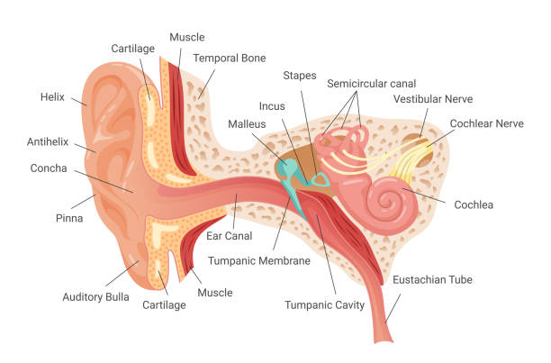
Structure of Ear and Their functions :-

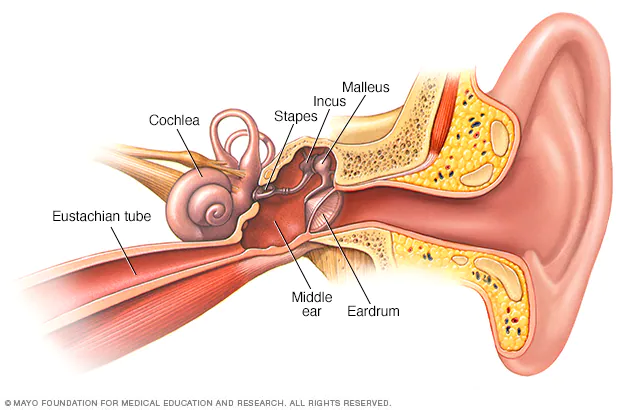
Human Ear are divided into Three part’s are following –

  1. Outer Ear ( Pinna , Ear Canal )
  2. Middle Ear ( Tympanium , Ossicle , Eustachian Tube )
  3. Inner Ear (Coachlea , 3 Semi-circular Canal’s)

Ear and Their functions
Ear

  • Stapes and Oval window are attached 

Outer Ear and their functions :-

  1. Pinna :- It’s work is collecting sound waves and transfer to the Ear Canal. The degine of Pinna is in this to collect the maximum waves of their surrounding area.
  2. Ear Canal :- 
    1. Hair :- The function of hair in Ear Canal is stop the foreign particle’s / dust particle’s that are present in Air.
    2. Wax (Cerumenous Glands) :- That is also work to trap the foreign particle’s / dust particle’s that are present in Air. 
    3. The work of the Ear Canal is conduct the sound wave’s.

Middle Ear and their functions :-

Middle Ear and their functions

  1. Tympanum :- It is made up of “Connective Tissue”.  It is vibrate’s when sound waves comes inside the Middle Ear.
  2. Eustachian Tube :- The Eustachian Tube connect Ear and Thrat and Pharynx. It is also maintain’s the air pressure balance occur’s tympanum.
  3. Ear Ossicle’s :- Smallest bones in body.
    • (3 Bone’s in Each Ear)
    • Ear Ossicles can done amplification of sound wave
    • The 3 Bones of Ear Ossicle’s are following
      1. Malleus
      2. Incus
      3. Stapes

Malleus :- Malleus is the largest & First bone in these three bones. It look likes Hammer in shape.

It is formed by modification of Articulate Bone.

Incus :-  Incus is the Middle bone in these three bones. It look likes Anvil in shape.

It is formed by modification of Quadrate Bone.

Stapes :- Stapes is the smallest & Last bone in these three bones. It look likes Stirrup in shape.

It is formed by modification of Hypo-Mandibular Bone.

Stapes is connected to cochlea through Oval(Ovalis) Window(Fenestra).

Window :-

They connect middle Ear ti Inner Ear. Their are two types are following :-

  1. Oval Window (Fenestra Ovalis) Ear and their functions
  2. Round Window (Fenestra Rotundus) Ear and their functions

1. Oval Window(Fenestra Ovalis) :-

The oval window, also known as the fenestra ovalis, is a connective tissue membrane located at the end of the middle ear and the beginning of the inner ear.

In the oval window the Scala vestibule (Perilymph fluid) are present

Helicotrema :- which connect the end part of , scala vestibuli and scalaa tympani.

The Reissnar’s Membrane are present in the Oval Window at the lower side of the Oval Window“.

And Oval window open in Scala Vestibuli.

2. Round Window(Fenestra Rotundus) :-

The round window , is also known as fenestra rotundus, is also a connective tissue membrane located at the beginning of the inner ear and the end of the middle ear.

The Basilar’s Membrane are present in the Round Window at the Upper side of the Round Window“.

The opening of Round Window in Scala Tympani. Ear and their functions

Note :- Ear and their functions Ear and their functions Ear and their functions 

  1. On Basilar Membrane the Techorial membrane was present and the Sterocilia(When the Sterocilia cell’s move then impulse will be generated now this impulse will send to the Brain) was present on the Organ of Corti (it is a unit of Hearing).
  2. The Endolymph fluid was present between the Oval window and Round window.

Sterocilia → Movement → Impulse generated → Signal Send to Brain .

Inner Ear and their functions :-

In the inner ear, the sound waves are converted into electrical energy, which your hearing nerve delivers to your brain as sound, making it possible for you to hear. At the same time, your inner ear monitors your movements, alerting your brain to changes so your brain can let your body know what to do to stay balanced.

Coachlea :-


The cochlea is a fluid-filled, spiral-shaped cavity found in the inner ear that plays a vital role in the sense of hearing and participates in the process of auditory transduction. Sound waves are transduced into electrical impulses that the brain can interpret as individual sound frequencies. Ear and their functions

Mechansim of Hearing :-

  • Amplified vibration enter’s into Scala vestibuli and they go from Scala vestibuli to Scala tympani because perilymph fluid also start vibrating but when perilymph  fluid was vibrating then both Reissnar’s and Basilar membrane will also start vibrating.
  • That vibrating conducted into scala media when vibrating enter’s into scala media then Endolymph fluid will also start vibrating.
  • When Endolymph was start vibrating then the stereocilia was also start vibrating / moving.   {Sterocilia is a cilia have a self movement generate. they can’t required any kind of external force for movement.}{Kinetocilia is a cilia they can’t required any kind of external force for movement. }
  • Sterocilia can generate signal / impulse and that signal / impulse willl send to the brain by the help of auditory nerve (it is the 8th cranial nerve)(cranial nerve is sensory in nature.)
  • Ear was located in temporal Bone.

Mechanism of Balancing :-

Responsible for balancing is Vestibular Apparatus.

Vestibular Apparatus 

  1. 3 Semi – Circular Canal → Macula →Dynamic Equilibrium [Ampulla , Macula , Cristae Ampullaris.]
  2. Otoliths / Otaconia / Ear Duct → Saccule & Utricle → Static Equilibrium

Structure of medula

  • In Cupula the jelly like fluid are present an fluid can help in balancing (Dynamic Balancing.)
  • Balancing jalantnus fluid was moving / vibrating then the stereocilia was also moving / vibrating and then generated signal and that signal was send to brain through neuron’s and the brain again send the signal to the muscle for movement.
  • All semi-circular canal’s are perpendicular to each other.

Saccule And Utricle :-

  • Stereocilia is responsible for static Equilibrium.
  • When we are standing start then the fluid will be parallel when we start bending one side then the fluid come’s one side and the fluid apply the pressure on stereocilia and then the stereocilia generate impulse and that impulse send to the brin and the brain again send signal’s to the body part’s and muscle to maintain the body balance 
  • so the Saccule and utricle are responsible for maintain the body balance.

What is Kidney and their function or process

Definition of Kidney/Excretory System :-

Kidney / Excretion system is a process of waste removal in the form produced during metabolic activities.


Kidney/Excretory Product’s :-

  1. Ammonia
  2. Urea
  3. Uric Acid
  4. Creatinine
  5. Guana 
  6. Hippuric Acid
  7. Greatine
  8. T.M.A.O.(Trimethyl/Amune Oxide)

Type’s of Organsim :-

  1. Ammonotelic :-
    • NH3
    • Most Toxic
    • Max H2O Needed
    • Readily Soluble in H2O
    • Ammonotelism
  2. Ureotelic :-
    • Urea moderately toxic 
    • Moderately H2O
    • Teretial Amptiuans
    • Example :-  Marune / Fishes
  3. Uricotelic :-
    • Example :- Bird’s , Reptiles, Land snail’s , Land Insect’s.

Human Excretory System :-

  1. A pair of Kidney
  2. A pair of Ureter’s 
  3. Urinary Bladder
  4. Urethra

{Right side kidney is slightly below left side due to above lying liver.}

So… What does the Urinary System Do ?

  • Keep my body balanced !
  • Cleaning the blood (metabolic waste’s N2)
  • Elimination of nitrogenous waste’s.
  • Excess solute’s (Ionic concentration)
  • Excess water (Osmoregulation)

Flow of urine :-

Renal Pyramid’s → Renal Papillae → Minor Cycles → Major Cycles → Pelvis → Ureter → Urinary Bladder → Outside.

Nephrone :-

  • The functional unit of the kidney are called Nephron.
  • Approx 1 million nephron are present in each.

2 Parts of Nephron

  1. Glomerulus :Glomerulus of capillaries in Bowman’s Capsule.
  2. Renal Tubule :→ 
    1. Bowman’s Capsule
    2. P.C.T. (Proximal Convoluted Tubules)
    3. L.O.H. (Loop of Henle)
    4. D.C.T. (Distal Convoluted Tubules)

Bowman’s Capsule :-

Bowman’s capsule is a part of the nephron that forms a cup-like sack surrounding the glomerulus. Bowman’s capsule encloses a space called “Bowman’s space,” which represents the beginning of the urinary space and is contiguous with the proximal convoluted tubule of the nephron.

Golomerules + Bowman’s Capsule  = Renal Capsule / Malphigiar

Renal Tubule :-

The renal tubule is a portion of the nephron responsible for homeostasis of fluids and electrolytes in the body. A renal tubule consists of a proximal convoluted tubule, loop of Henle, distal convoluted tubule, and collecting duct.

Blood Supply :-

Heart → Aorta → Renal Artery →(enter Kidney) → Approx 1 million Afferent Arterioles → Glomerulus → Efferent Arteriole→ Peri-tubular (P.C.T. + D.C.T.) & Vasa Recta (L.O.H.) → Renal Venules →  Renal → Inferior venacava → Heart.

Types of Nephron :-

Their are 2 types of Nephron

  1. Cortical Nephron
  2. Juxtamedullary Nephron

1. Cortical-Nephrons(80%-85%) :-

  • Loop of Henle
  • No Vasa Recta
  • Major part lies in cortex region.
  • No role in H2O Conservation.
  • Peri-tubular capillaries well developed .

2. Juxta-Medullary Nephrons :-

  • Major role in H2O conservation 
  • Counter current Mechnism.

Physiology Of Excretion :-

  1. Urea Formation :- (Liver) {Liver cell → Hepatocytes → Mitrochondria or Cytoplasma} → {Urea Cycle / Kreb’s Hensleit cycle / Ornithine Cycle}
  2. Urine Formation :- (Kidney)

1. Urine Formation :-

The three steps of urine production are filtration, reabsorption, and secretion. In filtration, molecules and ions pass from the blood to the filtrate within the glomerulus. Reabsorption retains important molecules, glucose, amino acids, ions, and water back into the bloodstream.

  1. Ultra Filtration (Unbiosed Process).
  2. Re-Absorption.
  3. Tubular Secretion.
Ultra Filtration :-

  1. Filtration under High Pressure
  2. Occur’s in Glomerulus + Bowman’s Capsule = Renal Capsule / Malphigiar.
    1. Filtration Membrane 
      1. Endothelium Capillary
      2. Basement Membrane
      3. Podocytes
    2. Glomerular Filtrate
      1. Glucose
      2. H2O
      3. Hormone
      4. Slat’s 
      5. Uric Acid 
      6. Urea
      7. Mineral’s 
      8. Vitamin’s
  • Only Remain in Blood that is called Blood Cell’s & Protein’s.
  • Ultra Filtration is Unbased Process. {Because they remove both substance useful & non-useful substances}
  • 125 ml / minute of Glomerular Filtrate
  • 180{G.F.R. = Glomerular Filtration Rate} ml / day of Glomerular Filtration

Net Filtration Pressure :- Net / Effective Pressure at which Ultra Filtration occur = 10 – 20 mm of Hg {(B.P. Blood Pressure = Increase’s B.P.) , (N.F.P. Net Filtration Rate = Increase N.F.P.)}

2. Re-Absorption :-

  • Re-Absorption is a Absorption of useful substance’s from Nephron Back to Blood. It occur’s in – P.C.T. , L.O.H. , D.C.T. , C.T.
  • Re-Absorption in P.C.T. & L.O.H. is known as Obligatory Re-Absorption.
  • Re-Absorption in D.C.T. & C.D. is known as Facultative Re-Absorption.

Active Re-Absorption :- Glucose , Sodium , Amino Acid.

Passive Re-Absorption :- Remaining Substance’s

  1. P.C.T.(Proximal Convoluted Tubule)
    1. Glucose = 100% 
    2. Amino = 100%
    3. Urea = 50%
    4. Sodium = 70%
    5. Potassium = 70%
    6. Phosphate = 70%
    7. Calcium = 70%
    8. Magnesium = 30%
    9. H2O = 70%
  2. Thick Ascending Limg(T.A.L.)
    1. Sodium = 25%
    2. Potassium = 20%
    3. Calcium = 25%
    4. Magnesium = 60%
  3. Distal Convoluted Tubule(D.C.T.)
    1. Sodium = 5%
    2. Calcium = 8%
    3. Magnesium = 5%
    4. H2O & Urea is Variable
  4. Collecting Duct
    1. Sodium =3%
    2. H2O & Urea is Variable

Counter Current Mechanism:- Done in 3 Level’s 

  • Between Descending L.O.H. & Ascending L.O.H.
  • Between Descending V.R. & Ascending V.R.
  • Between Adjacent Lemb’s of V.R. & L.O.H.

3. Tubular Secretion :-

  • Active Process
  • Blood Nephrone 
  • Only method of Blood Filtration in which Lack of Glomerule’s.

Excretion :- Filtration – Re-Absorption + Secretion.

Urine :-

95% of volume of Normal Urine is Due to Water.

Organic Component’s :- 

  • Urea
  • Urobilinogen
  • Uric Acid
  • Creatinine
  • Amino Acid
  • Metabolite’s Of Hormone’s

In-Organic Component’s :-

  • Cation’s – Na+ , K+ , Ca++ , 
  • Anion’s – Cl- , SO4- , HCO3- , HPO4+

Regulation Of Kidney Function’s :-

  1. A.D.H. :- Hypothalamus
  2. R.A.A.S. :- JGA (Juxta Glomerular Appaaratus)
  3. A.N.F. :- Heart

1. A.D.H.(Anti Diuretic Hormone) :-

  • Vasopressin (other name) Vessel Contriction
  • Diuresis (Excessive Urination)

Function’s :-

  1. Prevent Excessive Loss of Water in Urine.
  2. Increase Re-Absorption of Water in D.C.T. & C.T.
  3. Vesocontriction (Blood Pressure Increase)

A.D.H. Produced by Hypothalamus

A.D.H. Released by Post Pituitary. 

  • Secreted Under Following Situation
    • Water Loss – Excessive (Sweating , Diarrhoea)

Osmoreceptor’s in Body , Detcet :-

Osmolarity of Blood (During Summer)

When Osmolarity ↑ (Salt ↑) / H2O↓

Send signal to Hypothalamus

Hypothalamus produce A.D.H.

Effect D.C.T. + C.T.

H2O Re-Absorption Increase from D.C.T. + C.T.

Less Urine Formation

Osmolarity of Blood (During Winter)

H2O Level Normal

No A.D.H.(Anti Diuretic Hormone’s)

H2O Lost in Urine 

More Urination

2. R.A.A.S.(Renin Angiotension Aldosyerone System) :-

Renin :- Kidney

Angiotension :- Liver

Aldosyerone :- Adrenal cortexdd

RAAS is stimulated by → Low B.P. (G.F.R. is decrease)(Na+ is also decrease)

  • Juxta Glomerular Cell secrete Renin (When Na+ level decrease)
  • Macula Densa Cell detect Na+ level in blood .

When B.P. decrease (Na+)

↓(Hypotension)

Detected by Macula Densa

Stimulates J.G.C. to release RENIN

Angiotensinogen(In-Active) → Secreated by Liver

Angio-Tension 1 (Less Potent){Angio – Veso , Tension – Constrictor}

↓(Angiotension Converting)

Angio-Tension 2(Strong / More potent) Enzyme (Lungs) 

  1. Angio
    1. Blood Vessels → Increase Vasocontriction → B.P. Increase’s
    2. Adrenal Cortex → Aldosterone → Increase Na+ Re-Absorption from D.C.T. + C.T. → H2O Re-Absorption → Blood Volume Increase’s → Blood Pressure Increase’s.
    3. Post Pituitary

3. A.N.F. (Atrial Natriuretic Factor) :-

When B.P. Increase (Hypertension)

ANF Secreted by ↓ Artrial Wall’s

Heart / Atrial Muscles release A.N.F.


Suppress R.A.A.S.

Na+ & H2O Loss in Urine 

B.P. Decrease’s (Diuresis) Excessive Urination.

Micturition :-

Practise of voiding / Emptying Bladder

Urine :- Slightly Acidic pH is 6.8 (Max. Capacity 350 – 500 ml)

150 ml Generate’s Feeling to void .        ⇒     Maximum Full Generate Strong urge to Void.

Micturition Reflex :-

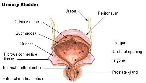
  • In Urinary Bladder stretch receptor’s are present they will send signal to brain through the sensory neuron.
  • At this time Detrusor muscle’s will be contraction.
  • Stretch receptor’s are present in the wall’s of Bladder.
  • Brain send message / signal to Detrussor Muscle’s and Uretheral sphincter’s for Void the Urine.

Ureter’s & Urinary Bladder :-

Kidney Bladder
Kidney Bladder

The urinary bladder is a hollow, spherical-shaped organ that holds urine (pee). For most people, it can hold 500-700 mL (about two cups) of pee. When you need to use the restroom, muscles in your bladder contract (tighten) and sphincter muscles in your urethra relax, allowing pee to flow out of your body.

Role of Kidney in Blood Formation :-

When Hypoxia occurs (Pressence Of O2 decrease’s in Air) {∴ At Higher Altitude the Count of R.B.C. will as Increased}

Stimulate’s Kidney to Produce Erythropoetin. {It required at leats 3-6 month 4-5 → 8 million/mm3}

                                    ↓

Stimulated Red Bone Marrow 

Increase R.B.C.(Red Blood Cell’s) Production.

Congestive Heart Failure (Part :- 1)

Defintion of Heart :-

The hollow, muscular organ that pumps blood through the body of a vertebrate animal by contracting and relaxing. In humans and other mammals, it has four chambers, consisting of two atria and two ventricles.

External Structure of Heart :-

  • The  Atrium can obtain 1/3 part. 
  • The Ventricle can obtain 2/3 part .
  • The Atrium and Ventricle are divided by Auriculo Ventricular Groove.
  • The Upper side of the Heart (1/3 part) can be divided by Inter Auricular Sulcus / Septum.
  • The Lower side (2/3 part) can be divided by Inter Ventricular Sulcus / Septum.

Protective Covering :-

It is Protected by Pericardium Layer.

  • Peri mean’s outer side .
  • Cardium mean’s heart .
  • Portective sac of connective tissue.
  • Filled with fluid.
  • Pericardium layer made of connective tissue.
  1. Fluid Work :- 
    1. Prevent from friction.
    2. Shock Absorbed.
    3. Prevent from Dehydration.

Internal Structure :-

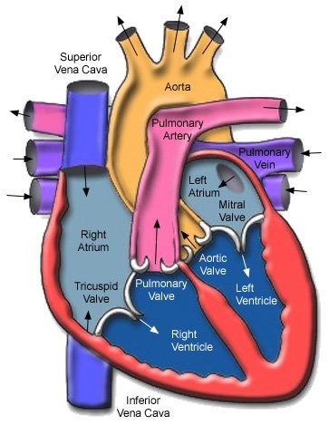
The De-oxygenated blood can be collected by superior Vena-cava into the Right  Atrium and then blood can be transfer into Right  Ventricle after full fill the Right Ventricular the blood can be transfer to the lung’s by the help of Pulmonary Artery and the blood can be oxygenated by the help of lung’s then the transfer blood again into the Left Atrium after full-fill the Left Atrium the blood goes to Left Ventricular chamber and after full fill Left Ventricular Blood can be transfer to the all body part’s by the help of Aorta.

Wall’s :-

Their are 3 layer’s of Heart

  1. Epi-cardium Layer
  2. Myocardium Layer
  3. Endo-cardium Layer

The wall’s are made up of 

  1. Volentry Muscle’s
  2. Involentry Muscle’s
  3. Cardium Muscle’s
  • The wall’s of Auricle’s is always thinner than the Ventricles 
  • Because Ventricles can pump the blood in the whole body part’s and the Auricle’s can collect the blood from all body.
  • When we supply the blood to whole body then the pressure will be high then it is compulsory that the wall of Ventricles is thicker than the wall of Auricle’s to absorb the pressure.
  • When we send blood to all body the we need more pressure do the wall’s of blood Ventricles thicker than the Auricle’s.
  • The thickness of the wall always depend’s on the Myocardium layer of the Heart.


Fossa Ovalis :-

The Fossa Ovalis is a hole which is present after the birth. 

Septum :-

The septum is the membrane which separate the right and left part of the heart . Separate De-oxygenated blood to Oxygenated blood.

Muscle’s of Heart :-

Ridge :-

Increase the surface area to hold blood in chamber’s.

  • Small ridge in atria (Pectinate Muscle in Atria)
  • Large ridge in Ventricle (Columnae Cornea/Trabeculae Cornea)

Chordea Tendineae :-

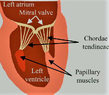
The chordae tendinae (CT) are strong, fibrous connections between the valve leaflets and the papillary muscles. These are attached to the leaflets on to the ventricular side and prevent the cusps from swinging back into the atrial cavity during systole.

Chordea made up off collagenHeart

Papillary Muscles :-

There are 5 papillary muscles in the heart originating from the ventricular walls. These muscles attach to the tricuspid and mitral valve leaflets via the chordae tendineae and functionally prevent regurgitation of ventricular blood via tensile strength by preventing prolapse or inversion of the valves during systole.

Value’s :-

Values are muscular flapes . Flapes are prevent from flow of blood in last chamber of the heart.

The 4 heart valves include the following:
  • Tricuspid Valve. Located between the right atrium and the right ventricle.
  • Pulmonary Valve(Pulmo-Semilunar Valve {P.S.L.V.}). Located between the right ventricle and the pulmonary artery.
  • Mitral Valve / Bicuspid Valve. Located between the left atrium and the left ventricle.
  • Aortic Valve (Aortic-Semilunar Valves{A.S.L.V.}).


Artery :- Artery they can take blood away from the Heart.

Vein / Venticle :- Vein / Ventrical they will collect the blood.

Structure of Heart :-


Structure of Heart

  • The De-oxygenated Blood can be collected in Right Atrium after full fill the Right Atrium the blood can be transfer into Right Ventricle after full fill the Right Ventricle then the blood transfer into Lunge’s through the Pulmonary Artery 
  • The lunge’s can filter the blood by the help of Alveli 
    1. In the Alveli the CO2 come out from Blood and O2 enter’s into the Blood this process is known as Gaseous Exchange 
  • After Gaseous Exchange the Blood come back again into the Heart in Left Atrium after the full fill the Left Atrium the Blood can transfer into Left ventricle 
  • After the full fill the Left Ventricle the blood can transfer into all body part’s through the Aorta. 

Working Of Heart :-

  • Our Heart is Myogenic {Myo – Muscle , Genic – Production}
  • Our Cardiac System can generate our beat by own/itself. The impales is produced within the Heart.
  • Our heart is Auto – Rythmicity.
  • In Human Cardiac System the SAN(Sino-Auricular Node) and the AVN(Atrio-Ventricular Node)
  • The SAN can take signal through Nerves from Brain and then the signal can transfer from SAN to A.V. Nodes and after that the electrical signal can transfer from A.V. Node to A.V. Bundles.
  • From A.V. Bundles the electric impales can transfer into Left and Right Branch of A.V. Bundles.
  • From Left to Right Branch of A.V. Bundles the electric impales can transfer into Purkinje Fibre’s.

S.A. Nodes :-

  • S.A.N. is a pacemaker 
  • S.A.N can produce impales 
  • Both Left and Right Artium can contract together and both Left and Right Ventricle are also contract together. 
  • S.A. Node is Located in Right wall of Right Atrium.

A.V. Nodes :-

  • A.V. Node is a Pacesetter.
  • A.V. Node is Located in Left wall of Right Atrium.
  • A.V. Nodes can re-amplify the impales and then the impales can transfer into A.V. Bundles.
  • The A.V. Bundles are divided into two branches.
    1. Purkinje Fibre {One can make network of fibre}
    2. The action potential from the S.A. Node reach the Ventricles in the following path.
  • Node ⇒ Atrial Muscles ⇒ A.V. Node ⇒ A.V. Bundle’s ⇒ Purkinji Fibre’s ⇒ Muscle’s of Ventricles (Wall of Ventricles)

Cardiac Cycle :-

Set of event’s which follow impales production by S.A. Node .

  • Relax :- Diastole.
  • Contraction :- Systole 
  • S.A. Node :(0.1 Second)- A.V. Node
  • Joint Diastole :-(0.4 Second)

  • Blood always flow from +ve pressure to -ve pressure {+ve means high pressure & -ve pressure }
  • In joint diastole all the chamber are relaxed so inside the Heart -ve pressure will be present and blood collected / stored in the Heart
  • T.V. & B.V. open & blood felling into auricles ⇒ Ventricle • 70% Ventricles are filled(Slow) at this time 

Atrial Systole(0.1 second) :-

  • After 0.4 second joint diastole the S.A. Node can generate impulse then the Auricles are contract and the blood can flow force fully into the Ventricles and the remaining (30%) part of the Ventricles are also filled with Blood. 
  • This can happened only in 0.1 second 
  • During this +ve pressure was present in auricle and -ve pressure present in Ventricles.
  • This can be done Rapidly.
  • When the Auricle are filling with Blood then that time the Ventricles will be relaxed.

Ventriclar Systole :-

  • In the Ventricles +ve pressure will be present.
  • I.V. and B.V. close.
  • After filled with blood the blood can apply pressure P.S.L.U.(Semi-Lunar valves) and A.S.L.V. and the blood go outside through the A.S.L.V. to the body.

                                                              Blood Goes Out

  1. Aort. → (oxygenated) →  Body
  2. Pulmonary Artery →(de-oxygenated) → Lunges
  • When the blood go outside Ventricles then the Ventricles then the Ventricles are fully empty & then the T.V. & B.V. can suddenly open and due to this all blood of Auricles Chamber’s are collect that blood can full down into the ventricles.

Cardiac Output :-

  • The volume of blood pumped out by each Ventricle , for each heartbeat , is known as the Stroke Volume.
  • The Volume of blood pumped out by the heart from each Ventricle per minute is termed Cardiac output
    • Per beat = 70 ml
    • Cardiac Output = S.V. × H.R.
    • Cardiac Output = 70 × 72
    •                            = 5040 ml/minute
  • Athletes have the ability to increase the stroke volume and cardiac Output.
  • The Cardiac Output generally increase in the active state and Decrease in resting condition.

Heart Beat :-

  • Number of times it beat’s/mint
  • Normal values = 70 – 80  times/mint
    1. Tachycardia = Heart Rate increase
    2. Bradycardia = Heart Rate decrease

Factors that Effect the Heart Beat :-

  1. Physical Activity
  2. Physical Condition & Health Condition.
  3. State of Mind
  4. Diet
  5. B.M.R.(Based Metaboli Rate)

Regulation of Heart Beat :-

  1. Nervous :- Medula Oblongata (Control)→ Cardiac centre (Send Message)→ S.A. Node
  2. Endocrine :- 
    1. Acetylcholine → P.A.N.S( Parasentatic Nervous System) → decrease (Heart Rate) 
    2. Thyroxin (Increase Heart Rate)
    3. Adrenalin
    4. Nor-Adrenalin 
      • Nor-Adrenalin and Adrenalin both are S.A.N.S. they Increase the Heart Rate 
    • P.A.N.S.(Parasentatic Nervous System) = They work in normal condition → Decrease Heart Reat
    • S.A.N.S.(Sentatic Autonomic Nervous System) = They work in emergency condition.

Blood Pressure :-

Pressure exereted by blood on walls of blood vessels.

  • B.P.(Blood Pressure) = S.P.(Systolic Pressure)/D.P.(Diastolic pressure)
      • Normal Systolic Pressure = 120 mm/Hg
      • Normal Diastolic pressure = 80 mm/Hg
    • Normal B.P.(Blood Pressure) = 120/80 mm/Hg
      • Hyper Tension (High B.P.) = 140/90 mm/Hg
      • Hypo Tension (Low B.P.) = 100/60 mm/Hg
    • Pulse Pressure = S.P. – D.P. ⇒ 120 – 80 =40mm/Hg
      • Blood Pressure measured by Sphygmomanameter (SPO2)
      • Heart Rate measured by Stethoscope

Blood Vessels :-

Artery :- 

Which carry blood away from Heart and toward’s the organs.

  • Tunica Externa made up of white fibrous connective tissue
  • Tunica Media :- Smooth muscles

Vein :

Which carry blood toward’s the Heart and collect from the organ’s & tissue’s.

  • Tunica interna :- Squamous Epithelium

Capillary :-
  • Tunica Interna & Tunica Media both are absent 
  • Tunica interna is present & it is also known as (Endothelium)

Lympth :-

  • Blood – R.B.C.(Red Blood Cell’s) – Platelets
  • Plasma + W.B.C.(White Blood Cell’s)
  • Plasma + Lymphocyte + Monocytes
  1. Lympth is colourless. 
  2. Proteins is also low.
  3. W.B.C. = 500 /nm3
  4. No Respiration Pigment 
  5. Important Role in defence against disease.
  6. If lymph flow around tissue that , it is also known as Interstial Fluid / Tissue fluid. 
  7. Acts as a middle man.
  8. Help in fat absorption.

Lymphatic Circulation :-

  • Lymph Capillary are belind/close at one side.

Diapedsis :-

Squeezing / coming out of W.B.C. + Plasma.

  • Heart → Aorta → Artery → Blood Capillary → Lymph vessels (Values more than veins) → Lymphatic Ducts (2 Lymphatic ducts are present at nack ression) → Sub Clavian veins (Right & Left) → Venacava → Heart.

What is Eye and their work and eye test (2024)

EYE :-

The postion of Human Eye’s in Frontal in Location.

Binocular vision can be done by human. (Binocular vision means we can see easily one object from both Eye’s in one time.)

eye
Eye’s

  1. These are the most sensory part of our body.
  2. These are protected by :-
    1.  Orbit(Bones protection{by the help of 8 type of bone’s})
    2. Eye’s Lashes [Upper Lashes(moveable) and Lower Lashes(inmoveable)]
    3. Conjuctiva 
      1. Eye Infection
        1. Pink Eye’s 
        2. Eye flew
        3. Conjuctivitie’s
    4. 3rd Eye lid (Nictitating membrane and Plicae Semilunaris) is vestigel [not functioning]

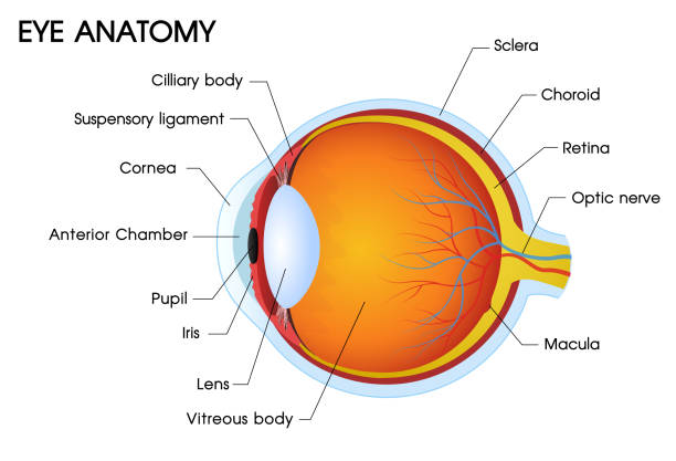
Structure of Human Eye :-

Eye
Eye

Hollow boll and filled with fluid

1. Made up of 3 Layer’s

    1. First layer/Outer Layer is Tunica Fibrosa.
    2. Second layer
    3. Third Layer is Tunica Nervosa 

Tunica Fibrosa :-

Tunica Fibrosa [1. Sclera{Backside(5/6)}  2. Cornea{Frontside(1/6)}]

[Note:-Cornea Get oxygen direct from atmosphere]

. Tunica fibrosa is help to maintain the shape of the Eye

Sclera can make Posterior part is 5/6 part.(White Par)

. Cornea can make Anterior part and they can make 1/6 part and they will be transparent.(The most successfull transplant cornea because their is no blood supply in cornea.)

Second Layer :-

It is made up of Blue Colour.

This is second layer and middle layer of Eye this is called Tunica pigmentosa and Tunica vascular.

Melanin pigment was present .

Melanin can absorb the extra light that was enter’s in the Eye.

Pigmentosa can also divided into 3 part’s.

  1. Choroid
  2. Ciliary Body
    1. Ciliary processes
    2. Ciliary Muscle’s
  3. Iris (Coloured part of the Human Eye)

The gap between two Iris is known as pupil.

Through the pupil light can enter’s into the eye Pupil’s size can regulated by the Iris. 

Third Layer :-

This is known as Tunica NervosaNeurosensory / Retina.

In this layer neuron’s are present. 

In this layer 3 Cell’s are present 

  1. Ganglionic 
  2. Bipolar 
  3. Photo receptor Cell’s 
    1. Rode’s 
    2. Cone’s 

Yellow spot (made up of photo receptor’s Cell’s ) was present just behind the pupil at the centre and the other name of Yellow Spot is Mocula Lukia.

The arrangement of the cell’s are Ganglionic then Bipolar at middle and the photo receptor Cell’s are present at last. 

At the yellow spot the ganglonic and bipolar Cell’s are absent.

Photo receptor’s Cell’s are two type’s

  1. Rode’s (Rode’s are used in night time / dimness)
  2. Cone’s (Cone’s are used in day time in brightness)

Len’s :-

The len’s are present just behind the iris.

The len’s will be connected by suspensory ligament’s.

By the help suspensory ligament can change the shape and size.

Ciliary Muscle and suspensory ligament can change the focal length of len’s.

Blind Spot :-

Blind spot is that place from that place the nerves are present / rise.

These nerve are known as Optic Nerve. 

Optic nerve is the Second cranial nerve.

That is sensory in Nature. 

Rode’s and cone’s both are absent on blind spot .

Between len’s and cornea the fluid was present that is known as Aqueous(Fluid) Humor’s(Watery).

Aqueous Humor’s was secrated by ciliary processes

Aqueous Humor’s is responsible for maintain the pressure in the Eye.

Behind the len’s the fluid is also present that is known as Vitreous(Jelly type) Humor’s(Watery)

Difference between Rod Cell’s and Cone Cell’s :-

Rod Cell’s :-

  • Cylindrically-shaped cell’s in the retina that respond to dim Light.
  • Outer sigment is rod Shaped.
  • Comparatively long.
  • Contain’s approximately 90 million’s Cell’s.
  • Distributes through the Retina.
  • Involved in peripheral vision. 
  • Only a single type occur in the retina. 
  • Responsible for non-chromatic vision.
  • Contain Rhodopsin 
  • More sensitive and responsible for night vision. 
  • Several Rod Cell’s connect to a single bipolar Cell’s.
  • Possess a poor acuity(Sharpness.)
  • Regenerative power is high.

Cone Cell’s :-

  • Cone-Shaped , visual receptor cell’s in the retina that are sensitive to bright light and colour.
  • Outer segment is Cone-Shaped.
  • Comparatively Long.
  • Contain approximately 6 million cell’s.
  • Mainly occur in the fovea
  • Can only tetct the image in the centre of the retina.
  • Three types occure in the retina 
  • Responsible for coloured vision.
  • Contain photopsin /Iodopsin.
  • Have a poor sensitivity and require bright light for vision.
  • One Cone Cell’s connected to one bipolar cell
  • Possess a higher acuity
  • Regenerative power is low.

Mechanism Of Vision :-

Mechanism of vision occur’s in two part’s 

  1. Focusing Part.
  2. Biochemical Part.

Focusing Part :-

  • Light must focus on Yellow Spot. 
  • Refraction Conjuctiva.
  •                  ⇓
  • Cornea (maximum refraction).
  •                  ⇓
  • Aqueous Humous.
  •                  
  • Len’s .
  •                  
  • Vitreous Humous.
  •                
  • Yellow Spot.

Part’s Involved :-

  • Len’s (Thinner)
  • Suspensory Ligament’s(Contract)
  • Ciliary Muscle’s (Relax)
  • Iris(Contrict)
  • Pupil(When Dilate then more light Enter.)

Biochemical Part :-

When fall’s on yellow spot which contain PRC (Cones)

                                 ⇓

Contain Visual → 1)Iodopsin(visual voilet) → Retinal + Photopsin → Red

                                                                                                               → Green

                                                                                                                →Blue

Red + Green + Blue = Equal Stimulation of all 3 cone’s can create White sensation

Pigments(photo-sensitive) → 2)Rhodopsin(visual purple) → Retinal + Scotopsin 

Undergo Biochemical Change’s

On moving from  bright light to dim light :-
  • We suffer from temporary blindness
  • Bright light has degenerated Rhodopsin(Bleaching)
  • For dim light → we require Rhodopsin. 
  • Rhodopsin ⇌ Retinal + Scotopsin

What is Ear and Their work (2024)

EAR :-

The engineering marvel because its sensory receptor can transduce sound vibrations with amplitude as small as diameter of atom of gold into electrical signal thousand times faster than photoreceptor can respond to light. It also contains receptor for equilibrium. It can also done Balance mechanisms of human body.


Ear
Ear

The Ear is divided into 3 main region :-

Ear
Ear

  1. External ear :- It collects sound waves and channel them inward.
  2. Middle ear :- It conveys sound waves to oval window.
  3. Internal ear :- It houses the receptor for equilibrium and hearing.

The external and outer ear consist of :–

a. Auricle
b. Auditory canal
c. Ear drum

Auricle (pinna) :-

Is a flap of elastic cartilage shaped like flared end of trumpet & covered by the skin
 The rim of auricle is k/a helix and inferior portion is lobule
 The lobule is highly blood supplied part of the ear
 The auditory canal is & curved tube about 2.5 cm long
 It is located in temporal bone & leads to ear drum
 The tympanic membrane or ear drum is a thin semitransparent partition between the auditory canal and middle ear
 The tympanic membrane is covered by epidermis and lined by simple cuboidal epithelium
 Between the epithelial layer is connective tissue composed of collagen fibers and fibroblast
 Near the exterior opening of auditory canal contains a few hair and specialized sweat glands known as cerumionous gland
 Ceruminous gland secrets cerumen or wax
 The combination of cerumen and hairs prevents dust and foreign object from entering the ear
 Cerumen also prevent the damage to delicate skin of external ear canal by water and insects

Middle ear :-

Ear
Middle Ear

It is the smallest filled cavity in the petrous portion of temporal bone
 It is lined by epithelium and separated by tympanic membrane from outer ear
 It forms the internal ear by a thin bony partition that contains 2 membrane covered opening oval window & round window
 3 smallest bone (Auditory ossicles) attached to it by ligaments which are connected by synovial joint

Auditory ossicles are :-

Malleus :-

It is a hammer shaped bone that attach to the internal surface of tympanic membrane (handle part). The head of malleus is articulate with the body of incus .

Inus :-

It is the anvil shaped bone and its head articulate with the head of stapes.

Stapes :-

It is stirrup shaped bone the footplate or base of stapes fits into oval window
Below the oval window there is another opening is round window which is enclosed by tympanic membrane .

2 skeletal muscles attach to the auditory ossicles :-
a) Tensor tympani :-

Supplied by mandibular branch of Trigeminal nerve (V)
It limits the movement and increase the tension on ear drum.

b) Stapedius :-

Supplied by the facial nerve (VII). It is smallest skeletal
Muscle. It protects the oval window and decrease sensitivity of hearing.

  1. Anatomy: The stapedius muscle is a tiny, cylindrical muscle located within the middle ear. It originates from the pyramidal eminence, a bony prominence on the posterior wall of the middle ear cavity, and inserts onto the neck of the stapes bone, one of the ossicles (small bones) of the middle ear.
  2. Innervation: The stapedius muscle is innervated by the facial nerve (cranial nerve VII). The motor fibers that control the stapedius muscle originate from the facial nerve nucleus in the brainstem and travel through the facial nerve before reaching the muscle.
  3. Function:
    • Dampening Sound: The primary function of the stapedius muscle is to dampen excessive movement of the stapes bone in response to loud sounds. When activated, the stapedius muscle contracts and pulls the stapes bone away from the oval window, which connects the middle ear to the inner ear. This action reduces the transmission of sound vibrations to the inner ear, protecting it from damage caused by loud noises.
    • Tuning: The stapedius muscle also plays a role in the process of sound localization and frequency tuning by adjusting the mechanical properties of the middle ear system.
  4. Reflex: The contraction of the stapedius muscle in response to loud sounds is known as the acoustic reflex or stapedial reflex. This reflex is a protective mechanism that helps prevent damage to the delicate structures of the inner ear from exposure to excessively loud noises.
  5. Clinical Significance:
    • Hyperacusis: Dysfunction of the stapedius muscle can contribute to conditions like hyperacusis, where individuals experience increased sensitivity to normal environmental sounds.
    • Acoustic Reflex Testing: The function of the stapedius muscle can be assessed through acoustic reflex testing, which measures the reflexive contraction of the muscle in response to loud sounds. This test is often used in audiological evaluations to assess hearing function and diagnose certain auditory disorders.

The anterior wall of middle ear contains an opening that leads to eustechian tube (pharyngotympanic)
It is consist of bot bone and elastic cartilage and connects the middle ear with nasopharynx
It is closed at the medial end (pharynx)
During swallowing and yawning its open and allow to air to enter or leave to middle ear until the pressure in the middle ear equal to atmospheric pressure

Inner ear :-

It is also known as labyrinth because of its complicated series of canal.it consist of 2 main division

a) Outer bony labyrinth
b) Inner membranous labyrinth

The bony labyrinth is series of cavities in the petrous portion of temporal bone. It is divided into 3 areas –

a) Semicircular canal :-
b) Vestibule:-
c) Cochlea :-

Semicircular anal and vestibule both the contains receptor of equilibrium and cochlea contains the receptor of hearing
The bony labyrinth is lined with periosteum and contains perilymph and chemically similar to CSF
Membranous labyrinth is a series of epithelial sac and tube inside the bony labyrinth. The epithelial membrane contains endlolymph
The level of K+ Ions is high for an ECF and play the important role to generate the electrical signal

a) Semicircular canal :-

superiorly and posteriorly from the vestibule there are 2 semicircular canal each of which approx. right angles to the outer 2
Based on their position they named as anterior, posterior and lateral semicircular canal. The anterior and posterior canals are vertical and lateral one is horizontal
At the one end of each canal is swallowen enlargement known as ampulla
The portion of membranous labyrinth lie inside the bony semicircular canal are known as semicircular ducts. These ducts connects with utricle and saccule
The vestibular branch of VIII CN consist of ampullary , utricular and saccular nerves and responsible for equilibrium

b) Vestibule :- 

Is the oval central portion of the bony labyrinth. The membranous labyrinth in the vestibule consist of 2 sacs called utricle and saccule which are connected by a small duct.

c) Cochlea :-

is a snail shell bony spiral canal which is anterior to vestibule
It makes almost 3 turns around a central body core known as modiolus

Cochlea divided into 3 channels :–

a) Cochlear duct (scala media)
b) Scala vestibuli
c) Scala tympani

Cochlear duct is a continuation of membranous labyrinth into cochlea and filled with endolymph
The channel above the cochlear duct is scala vestibuli which ends at the oval window
The channel below is scala tymapni which ends at the round window
Both scala vestibuli and tympani are the part of bony labyrinth and Contains perilymph and both are completely separated to each other by scala media except helicoterma
Vestibular membranes separates the cochlear duct from scala tymapni
On the basilar membrane organ of Corti (Spiral Organ) Rests

Two groups of hair cells are presents-
a) Inner hair cells : arranged in single row
b) Outer hair cells : arranged in 3 layers

At the tip of hair cells 40-80 long hair like micro villi arranged known as stereo cilia
At the base end inner & outer hair cells synapse with VIII CN
A flexible gelatinous membrane cover the hair cells of spiral organ known as tectorial membrane. The body of hair cells rest on basilar membrane

PHYSIOLOGY OF HEARING :-

1. The auricles collects and directs the sound wave in auditory canal
2. Sound wave strike on tympanic membrane and the high & low pressure of air cause tympanic membrane to vibrate
3. The central area of ear drum connects to malleus which also start to vibrate. The vibrations is transmitted from the malleus to incus to stapes4. As stapes moves back & forth it pushes the membrane of oval window in & out
5. The oval window vibrates 20 times more vigorously than ear drum
6. The movement of oval window sets up fluid pressure waves in perilymph of cochlea
7. Oval window pushes on perilymph to scala vestibuli
8. Pressure waves are transmitted from scala vestibuli to scala tymapni to round window
9. Pressure wave push the vestibular membrane back & froth & creates the pressure waves in the endolymph inside the cochlear duct
10. The pressure wave in the endolymph causes the basilar membrane to vibrate
11. This moves the hair cells of spiral organ against tectorial membrane
12. This leads to bending of hair cells stercocilia which produce receptor potential that lead to generation of nerve impulse and leads to hearing

What is blood and their work (2024)

Blood and It’s Component’s :-

  • Total volume of blood is 5-6 Liter in human body.
  • The pH value of blood is 7.4 litre
  • Colour of blood in arterie’s = Bright Red
  • Colour of blood in vein’s =Dark Red

Plasma

Blood Contain’s

  •  55% Blood
  • White Blood cell’s and Platelet’s
  • Red Blood Cell’s

Plasma :-

Blood
Plasma

Definition :- Transport nutrient’s , hormones and proteins. It is a yellow liquid that makes up about 55% of the body’s blood volume. 

Plasma contain’s  Yellowish part of the blood

Plasma mainly contain’s 

  • H2O (90% – 92%)
  • Protein’s (6% – 9%)
  • Nutrient’s
  • Gases
  • Waste
  • Salt’s
  • Hormone’s
  • Heparin (anti – coagulant)
  • Coagulant Factor (inactive)

Type’s of Plasma :-

1. Albumin’s

2. Globulin’s 

3. Clotting (Fibrinogen)

Albumine :-

The albumine is maintain osmotic Balance of Blood.

Globulin’s :-

The other name of globulin is Immunoglobulin

The Immunoglobulin is also known as Antibodies.

Clotting Factor’s (Fibrinogen) :-

The clotting factor prevent excessive bleeding.

Formed Element’s :-

Plasma :- Transport nutrient’s , hormones and proteins. It is a yellow liquid that makes up about 55% of the body’s blood volume. 

Platelet’s :- Form clot’s to stop bleeding . Platelet’s make up less than 1% of blood.

Red Blood Cells :- Carry fresh oxygen through the body and remove CO2 (Carbon dioxide). RBC’s make up about 40% – 45%of blood.

White Blood Cells :- Part of the body’s  immune system , detect and fight viruses and bacteria. There are five major types of WBC’s and they make up less than 1% of blood.

Red Blood Cell’s :-

Red Blood Cell’s

It is also known as Erythrocytes

The Erythrocytes is divided into two part’s

The first is Erythro and the second is Cytes

  • The meaning of Erytro is Red 
  • The meaning of Cytes is Cell

The name of Erythrocytes is formed from Erythropoesis

  • The meaning of Erythropoesis is Red Bone Marrow.

Red Blood cell’s are started from Erythroblast . The meaning of blast precusor cell / mother cell

  • The formation / process of RBC is 
    • Erythroblast (Precursor cell)
    • Reticulocyte (Immature R.B.C.)
    • Mature R.B.C. is
      • Discoidal
      • Enucleated
      • Loss of organelles

The life spend of R.B.C. is 120 day’s .

After 120 day’s they goes in our graveyard of R.B.C. is spleen. Because after 120 day’s the R.B.C. was go into the spleen.

The Spleen is also known as Emergency blood blank of the body {in case of excessive bleeding spleen give RBC to the liver and liver divide the hemoglobin(Hb) }

Normal Value / amount / count in Male :- 5 to 5.5 million/mm^3

Female :- 4 to 4.5 million/mm^3

  • Abnormal condition :-
    • Number decreases :- Erythropenia / Anemia 
    • Number increases :- Polycythemia  

Transport of Gases (H.B.) O2:- Polycythemia 

CO2 :- Oxyheamoglobin

CO :- Carboxy (Hb)

White Blood Cell’s :-

White Blood Cell’s

(Soldier’s of body) 

Normal Count in W.B.C. = 6000 to 8000 / mm^3

Their are Mainly 2 types of WBC’s 

  • Granulocyte’s
    • Multilobed Nucles (Neutrophil)
    • Bilobed N/Essinophil (Acidophil)
    • S-Shaped (Basophil)
  • Agranulocyte’s
    • Monocyte’s (Largest WBC’s)
    • Lymphocyte’s
    •  (Smallest WBC)

Multilobed/Neutrophil :-

It is also known as “Poly morpho nuclear / Leucocyte (P.M.N.L.)”

  • The meaning of ‘Poly’ is “many” and the ‘morpho’ is “structure”
  • The meaning of ‘Leuco’ is “White” and the meaning of ‘cyte’ is “Cell’s”

Acidic and basic granules are present in this W.B.C.

  • Acidic show red in colour
  • Basic show blue in colour

Phagocytosis (they attack first on forein body particle’s).

Bilobed N / Eosinophil :-

  • It is also known as “acidophil”.
  • Acidic granule’s are present in this W.B.C.
    • The acidic granules are show red in colour.
  • They are active in allergies.

S-Shaped N/Basophil :-

It is also known as “Secretory”

Basic granules are present in this W.B.C.

  • Basic granules are shown blue in colour.
  • They secret –
    • Heparin (anticoagulant)
    • Histamine (Inflammation :- Pain , swalling , Redness )
    • Serotonin ( Anti – Inflammation)

Monocyte’s :-

It is the largest W.B.C.

The shape of the Nucleus just like Kidney.

In the Monocytes W.B.C. they contain Macrophages.

Lymphocyte’s :-

It is the smallest W.B.C. 

The Lymphocyte’s contain’s “Large Nucleus”

In the Lymphocytes the nucleus cover’s “Scanty cytoplasm”

The lymphocytes contain’s “Immune System (Anti-bodies)”

Abnormal Condition of W.B.C. Number’s :-

  • When Number of W.B.C. is increase’s is known as“Leucocytosis”
  • When Number of W.B.C. is decrease’s is known as“Leucopenia”
  • When Number of W.B.C. is more increase’s is known as “Leukaemia”
    • Leukaemia is also known as “Blood Cancer”

Platelet’s :-

The other name of Platelet’s is Thrombocytes 

  • The meaning of the Thrombocus is clot

The Thrombocytopoesis is drived from Red Bone Marrow 

  • Platelet’s are irregular
    • Cell fragment 
    • Enucleated
    • Sticky
    • Delicated

Abnormal Condition’s :-

  • When the Number are decrease’s :- Thrombocytopenia
  • When the Number are increase’s :- Thrombocytosis
Coagulation :-
  • Heparin (inactive on oxidation)
  • Formation of Permanent Clot Formation Process
    • Tissue injury
    • Platelet’s rapture or Aggregation of Platelet’s (This is temporary clot formation)
    • Release Thromoboplastic
    • Thrombokinase
      • Thrombokinase
        • Prothrombin(Inactive)
        • Thrombin(Active)
        • Fibrinogen
        • Fibrin
          • Fibrin :- It’s deals with Dead cell’s and it makes clot / Thrombus.

Blood Groups :-

Father of blood group is Karl Lardsteiner.

Blood group discovered in ‘1901’

Theory of Karl Lardsteiner’s :-

I have recently observed and started that the serum of normal people is capable of clumping the red cell’s of other healthy individuals . As commonly expressed, it can be said that in these cases at least two different kinds of agglutinins exit , one kind A, the other in B, both together in C. The cell’s are naturally insensitive to the agglutinins in their own serum.

Blood Group’s :-

Table

  • Blood group’s mainly depend’s upon Protein.
  • Antigen’s are present on the surface of R.B.C.
    • Their are mainly two types of Antigen’s 
      • ABO System
      • Rh System

A.B.O. System :-

Blood Group’s are

  • A
  • B
  • AB
  • O

Ag{Antigen’s} (R.B.C. Surface)

  • A
  • B
  • AB
  • None

Antibodies (AB) in Plasma 

  • Antibody – B  {Antigen – A}
  • Antibody – A {Antigen – B}
  • No Antibody {Antigen – AB}
  • Antibody A & B {Antigen – O}

Mismatching of Blood Group :-

When the blood group is mismatched then the blood cells will be destroy.

At the antigen (A) the antibody (B) are present 

  • Antigen O can contain both antigen’s (A and B) both are present .
  • Antigen O is universal Donar.

{Group A}  (Ab) = Antibody B , Ag(Antigen)=A , Donate to= group A & AB , Receive from= A&O

{Group B}  (Ab) = Antibody A , Ag(Antigen)=B , Donate to= group B & AB , Receive from= B&O

{Group A & B}  (Ab) = Antibody (None) , Ag(Antigen)=A&B , Donate to= group AB , Receive from= A, B, AB, and O(Universal Receiver)

{Group O } (Ab) = Antibody A&B , Ag(Antigen)=None , Donate to= group O, A, B, A & AB(Universal Donar) , Receive from= O.

Rh System :-

Rh Factor

Rh stands from “Rhesus Monkey” it is discovered from the Rhesus Monkey. 

If on the R.B.C. cell’s the Rh cell’s are present then we known as ‘Rh Positive’ if Rh cell’s are absent then we known as ‘Rh negative’.

Rh positive is known as donar Rh positive always gives both positive’s or negative’s (Rh) but Rh negative’s can only take’s from negative. 

  • An the Antigen’s Rh positive are present but Rh negative are absent.
  • In the Antibodies Rh positive are absent but Rh negative are present.
  • Rh positive can receive from Rh positive and Rh negative but Rh negative can receive from Rh negative.
  • Rh positive can receive from Rh positive and Rh negative but Rh negative can receive from Rh negative.
  • Rh positive can donate to the Rh positive and Rh negative can donate to the Rh positive and Rh negative both.

Rh incompatibility :-

{The Rh incompatibility always occurs between the mother and the baby after 1st pregnancy and they will be effect during 2nd after 2nd pregnancy.

They will be affect on the baby due to Rh incompatibility baby will be born Still dead.}

Normal Rh incompatibility

  • If Mother is Rh positive and Father is Rh positive then baby is also Rh positive.
  • If Mother is Rh negative and Father is also Rh negative then baby is also Rh negative.
  • If Mother is Rh positive and Father is Rh negative then baby is also Rh positive.

Abnormal Rh incompatibility

  • If Mother is Rh negative and Father is Rh positive then baby is also Rh positive

This condition leads to Erythroblastosis Foetalsis / Haemolytic Disease of Newborn (H.D.N.)

  • During the first pregnancy IgM will never cross the placenta because IgM size will be bigger.
  • During the second pregnancy IgG will cross the placenta because 
    • IgG destroy the foetal R.B.C.
      • When IgG destroy then Severe Anaemia and severe Jaundice can be done.

To prevent from IgG the injection will be given to the mother after the 1st delivery . So the injection (Rhogma) will create the brarrier for the IgG then the baby will be normal.


What is Internal Respiration Airway and how it is work (Part:-3)

Respiratory Airway/Respiration :-

Nose :-

It is first part of  respiratory air way that is located in between both orbits.

Anatomically Nose heaving two openings –

A) Anterior nasal Orifies / Porse :-

nasal cavity opens exterior through anterior nasal pores.

B) Posterior Nasal Pores :-

These are located posteriorly through while nasal cavity opens in nose pharynox posterior 

Respiration in NASAL SEPTUM :-

  1. It is a anatomical strcture that divide’s nasal cavity into two compartments right and left.
  2. Posterior half part of nasal septum is made of bone. 
  3. Anterior half part of nasal septum is made of elastic cartilage.

Mucus Membrane of Nose :-

  1. It lines nasal cavity.
  2. Mucus secrating cell are pre.in nasal mucosa. 
  3. Hair folids are pre. in nasal mucosa. 
  4. Para nasal cynes (funtal cynes, maxilary cynes, ethmoid cynes) opens in nasal cavity through in nasal mucosa.

Naso Lecrimal Duct :-

  1. It is a tubular strcature that aries from lecrimal sac and pre in letral wall of nose.
  2. It Opens into nasal cavity.
  3. Different bone that folds wall of nose are –  
    1. Nasal Bone’s
    2. Ethmoid Bone
    3. Maxilla Bone
    4. Nasal Concha
    5. Womer
Functions :-

1.Transportation:-  Air during inhalation and exhalation transmits transmits through nose.

2. Filtration :- Dust particles are traped nasal mucosed.

3. Humidification :- In healed air become most when it comes in contact with mucus. 

4. Warmification :- In healed , air becomes warm when it comes in contact with mucus membrane.

5. Resonence :- Nose provides resonance to voice to voice ( para nasal cynes P.N.C. )

6. Allfection :- Nerve ending of allfectory nerve are pre. in mucus membrane of nose.

Nerve ending stimulats when desolved small particles comes in contact.

As a result dipolerization occures and impulse  generate.

Generated nerve impulse conduct upto allfectory centres that are pre. in brain (ceribrum)

Sense of small occurs after inter pretation.

Trachea :-

  • This is a cartilaginous tube between the larynx and the lungs. It is a made of C is placed at the back and is covered by fibrous tissue.
  • The cartilage rings hold the trachea open for the passage of air.  The goblet cells secrete mucus.

Larynx :-

It is also known as voice box .

It is a part of respiratory air way that is pre. in mid axis of Neck.

Superiorly it is continous as pharynx while inferiorly its continous as trachea. 

Anatomical Relation :-

Anterior :- Skin , superficial fascie difascia.

Letarly :- Thyroid lobes and carotid verseles, nerve fibers.

Superiorly pharynx, hyoid bone

Inferiorly :-

First tracheal cartilage wall of larynx:-

wall of laynx consist cartilages.

Cartilages are inter connected with the help of ligaments are –

Unpaired cartilage

  1. Epiglotis
  2.  Thyroid
  3. Cricoide

Paired cartilage

  1. Arytenoid cartilage
  2. Corniculate cartilage
  3. Cunieform cartilage

1. Nine cartilages are pre. in wall of larynx.

2. Epiglotis is a elastic cartilage 

3. It is a lid like structure that closes in-large of larynx during deglutition (swalowing) 

4. Thyroid cartilage having two plates.

5. Both plates form in accute angle in mid axis of Anterior wals of larynx.

6. This angle is more prominant in male and it is called adams apple.

7. Cricoad cartilage is a ring shaped cartilage that is pre. below to thyroid cartilage and above to first tracheal cartilage.

8. Arytenoid cartilage is a paired cartilage and pre. in posterior wall of larynx.

9. It is conical in shape .

10. Corniculate cartilage is a paired cartilage and pre. in posterior wall of larynx.

11. Couniform cartilage is also a paired cartilage and pre. in posterior wall of larynx.

Mucus Membran :-

1. It is inner most layer of larynx.

2. It lines larynx interiorly.

3. Ciliated columnor epithelial cells lines mucus membrane. Mucus secrating columnor cells.

4. Blood vessels, nerve fibers, limph vessels and lymphoid folids are fire is sub – mucosal resion.

5. Mucus membrane is modified and forms folds.

6. The fold of mucus membrane that are pre. in inlate of larynx are called fowls vocal cord.

7. Another two folds of mucus membrane pre. below to fowls vocal cord are called true vocal cord.

8. True vocal cord are arranged in ‘V’ shaped. 

9. Anteriorly vocal cords are attached with a thyroid cartilage.

10. Posteriorly attached with arytenoid cartilage. Vocal cords are related to voice production. 

11. A space that is pre. in between true vocal cord is called rimoglotis.

Blood Supply :-

Arterial supply vein – laringial artry , laringial vein

Nerve supply – recrunt laringial nerve.

Functions :-

1. It transports air during inhalation and exhalation .

2. Humidification

3. Warmification

4. Filtration

5. Phagasytosis:- Tymphoi[ez-toc]d folids form antibodies and lymphocyte cells that degrate micraps.

Voice Production :-

Vocal cords are pre. in larynx that vibrats during forcefull flow of air and voice production accure high pitch voice production occures in female as more cords.

– Lord voice production or low pitch voice in male is due to low tendency in vocal cord.

Trachea :-

Defination :-

It is a wind pipe that pre. in mid axis of neck and in superior or media sternum.

It is a tubular structure that is pre below to larynx and runs down word up to cornia.

Length :-

It’s normal length is 10.5 cm

Anatomical Relations :-

Anterior :- Skin , superfacial fecia and deep facio

Laterly :- curotid vessels 

Superiorly :- Larynx

Inferiorly :- Bronchus

Posterirly :- Essophagus

Blood Supply :-

Arterial :- 

1. Branches of inferior thyroid artery 

2. Lower part is supplied by Branches of Bronchia artery

Veinus Collection :- 

1. Vein yellow artery

2. Nerve Supply – Autonomic nerve intervent.

Histology :-

Histologicaly different layers are pre. in it’s wall

A) Fiberus Layer (outer) :- 

It is made of fibrous connective tissue

B) Cartilagenus Layer :-

C – shaped cartilages are pre. in this layer 

Skin area of cartilage is directed posteriorly.

Tracheal muscles is pre. in skin area.

C) Sub-mucosal layer :- 

It is made of arealor connective tissue.

Blood vessels, nerve vessels , nerve fiberus , lymphoid fluids are also pre. in this layer.

D) Mucus membrane :-

It is inner most layer that is lined up by ciliated columnar epithelial cells 

Mucus secrating cells are pre.

Function:-
  1. Transportation of air 
  2. Humidification
  3. Warmification
  4. Filtration
  5. Phagacytosis
Bronchus :-


Defination:- It is a part of a respiration airway that are pre. in medisternum. There are two branches – Right and Left 

  • Both Branches aries from trachea
  • Terminal end of Bronchus enters in lung through root of lung .
  • Each Bronchus devides in secondary Bronchealus
  • Two in left side and three in right side.
Blood Supply :-

Broncheal Artery

Veinus Collection – Broncheal vein 

Nerve Supply – Autonomic nerve ineratis

Histology :-

A) Fiber’s layer(outer)

B) Cartilagenus Layer :-

Broncheal muscles are pre. in skip area.

C) Sub Mucosa

D) Mucus Membrane 

Function :-
  • Transportation
  • Filtration
  • Humidification
  • Phagocytosis
Broncheal Tree :-
  • Primary Bronchus inters in lung paranchyma through root of lung.
  • Primary Bronchus divide in secondary and tersory bronchus
  • Tersory bronchus ferther dicide 5-6 types.
  • As bronchus divide it’s lumane becomes narrow and size of cartilage becomes small.
  • Size of broncheal muscles becomes large.
  • At the level of respirotry broncheal cartilage is absent in it’s wall.
  • Broncheal muscle’s ancirkil wall of respiratory broncheal.
  • Respiratory bronchioles opens in alveolar duct and alveoli.

Alveoli :-

  • It is a terminal part of respiratory air way in which alveolar duct’s open.
  • It is conical in shaped.
  • It’s base is directed toward’s peripharilly while apex is directed towards root of lung.
  • Lung alveoli is lined up by squamus epithelial cells.

Also Read this

and this also

What is External respiration and how it is works(Part :-2)

Respiration & Breathing :-

BREATHING :-

Principal:- Air always moves from High Pressure to Low Pressure 

Their are two types of breathing :-

1.  Inhalation (Inspiration)

2.  Exhalation (Expiration)

Inhalation :-

Inhalation is represent (U,F,O) .

U:- Upward

F:- Forward

O:- Outward

The contraction of Inter coastal muscles will be done.

During Inhalation the Diaphragm was also play an important role in Breathing . During Inhalation the diaphragm will flate because they support to the Inhalation. Ribs and sternum raised and they represent (U.F.O.= Upward, Forward and Outward.)

When we Inhale then the Diaphragm will move downward because we want to increase volume inside the thoraic cavity

When we Inhale then the volume of Thoraic Cavity will be increase.
When we Inhale then the Pressure of Thoracic cavity will be decrease as compare to the outermost atmosphere.

Exhalation :-

IN the Exhalation is Ribs moves in the form of (D.B.I.)

D:- Downward

B:- Backward

I:- Inward

During Expiration the ribs and sternum moves/returned to original position and then the Volume of thorax decreased. 

When the Ribs and Sternum will become their original position/shape then the volume of thorax will be decreased and then the air expelled from lungs.

The inter coastal muscles will be relax when the Ribs come out their original position.

When we Exhale then the Diaphragm will move downward Ribs will move inward. Then the diaphragm becomes their original shape they become in the dome shape. When the diaphragm becomes dome shape then air come out side then we can easily Exhalation process and alveoli will contract during this process.


FORCEFUL BREATHING :-

Structure involves in forceful breathing.

  1. Ribs 
  2. External (Inter Coastal Muscles)
  3. Diaphragm
  4. Internal (Inter Coastal Muscles)
  5. Abdominal Muscles

PULMONARY VOLUMES AND CAPACITIES :-

1. Tidal Volume(T.V.):-    Volume of air inspired or expired during a normal  respiration(volume of air renewed in the respiratory system during each breathing).     500 ml   

2. Inspiratory reserve volume (I.R.V.) or complemental air :- Additional volume of air, that can inspire by forceful inspiration.  2500-3000 ml

3. Expiratory reserve volume(E.V.R.) or supplemental air :-  Additional volume of air, that can expire by  a forceful expiration.  1000-1100 ml     

4. Residal volume(R.V.) :-  Volume of air remaining in the lungs after a forcible expiration.   1100-1200 ml   

PULMONARY CAPACITIES :-

1. Inspiration Capacity (I.C.) [T.V.+I.R.V.] :- Volume of air that can inspire after a normal expiration.  3000-3500 ml volume 

2. Expiration Capacity (E.C.)[T.V.+E.R.V.] :- Volume of air that can expire after a normal inspiration. 1500 – 1600 ml volume

3. Functional Residual Capacity(F.R.C.) :- Volume of air that will remain in the lungs after a normal expiration [E.R.V.+R.V.]. 2100 – 2300 ml volume

4. Vital Capacity (V.C.)[E.R.V.+T.V.+I.R.V.] :- Maximum volume of air that can breath in after a forced expiration or maximum volume of air that can breath out after a forced inspiration. 3500 – 4500 ml volume

5. Total Lung Capacity(T.L.C.) :- Total volume of air lungs after a maximum inspiration.[R.V. + E.R.V. + T.V. + I.R.V. or V.C. + R.V.] 5000 – 6000 ml volume

REGULATION OF BREATHING/RESPIRATION :-

  1. Nervous:- The parts of brain which can help to breath 

1. Pons Varolhi:-  In the pons varolhi their are two parts :-

I).  Pneumotoxic Centre :- It is the switch off centre of the inhalation and inspiration . 

II). Apnestic Centre :- It is responsible for modulating the depth and rhythm of breathing.

2. Medulla Oblongata:- In the Medulla Oblongata is also two parts:-

 I). Dorsal Inspiratory Group :- They also control inspiration they control that how many time we will done inspiration.

II). Ventra Respiratory/Respiration Group :- They control both inspiration and expiration.


CHEMICAL :-     

Their are two chemical which can help to regulate the breathing 

  1.  CO2(Carbon Dioxide )
  2.  H+ Ions
  • When the number of CO2 ions is more and H+ ions is also more than the rate of breathing rate that will be also more.
  • When the number of CO2 ions is less and H+ ions is also less than the rate of breathing rate that will be also less.
  • If their is no CO2 molecules than their no breathing can be done.

MECHANICAL :-

( Hering – Breur’s Reflex)

    1. Inhalation
    2. Bronchi and Bronchioles Stretch
    3. Strech receptors in their wall’s send impulse to brain
    4. Brain send’s message to stop inhalation and start exhalation.


GASEOUS EXCHANGE/INTERNAL RESPIRATION :-

Gaseous Exchange mainly depend’s upon 

1. Pressure / concentration gradient ( High Pressure to Low Pressure).

   2. Solubility of gases.

3. Thickness of membranes.

4. Surface area of repiratory membrane.


GAS EXCHANGE BETWEEN ALVEOLI & BLOOD OR BLOOD TISSUE :-

ALVEOLI AND BLOOD  :- 

The gaseous exchange between Alveoli and blood the air gaseous exchange always moves from high pressure to low pressure 

  1. In Alveoli the pressure of oxygen  (O2) is 104 mm/hg and the pressure of carbon dioxide (Co2) in Alveoli is 40 mm/hg and the pressure of (O2) oxygen in Blood  is 40 mm/hg and the pressure of CO2 (carbon dioxide) in blood is 45 mm/hg 
  2. Now the air moves from High Pressure to Low Pressure means air moves from Alveoli to the Blood 
  3.  Now the pressure of oxygen (O2) in Blood is 95 mm/hg and the prssure of carbon dioxide (Co2) in blood is 40 mm/hg.

Respiration
Respiration

BLOOD & TISSUE :-

The gaseous exchange between Blood to Tissue the air gaseous exchange always moves from high pressure to low pressure 

  1. In Blood the pressure of oxygen  (O2) is 95 mm/hg and the pressure of carbon dioxide (Co2) in Tissue is 40 mm/hg and the pressure of (O2) oxygen in Tissue is 20 mm/hg and the pressure of CO2 (carbon dioxide) in Tissue is 45 mm/hg 
  2. Now the air moves from High Pressure to Low Pressure means air moves from Blood to the Tissue.
  3.  Now the pressure of oxygen (O2) in Tissue is depends on the tissue it’s range is (20 to40) mm/hg and the pressure of carbon dioxide (Co2) in Tissue is also depends on the Tissue the range will be (45 to 48) mm/hg .

Respiration
Respiration

RESPIRATION GASES                                          O2         CO2    

Attmospheric air (inspired air)                                                           159            0.3

Expired Air                                                                                            116             32

Alveoli Air                                                                                             104             40

Blood De-oxygenated                                                                          40               45

Blood Oxygenated                                                                               95               40

Tissue                                                                                                     40               45

What is Respiration and their work (Part:-1)

Respiration :-

The steps of respiration involved are the following:-

  1. Breathing or pulmonary ventilation by which atmospheric air is drawn in and CO2 alveolar air is released.
  2. Diffusion of gases ( O2 and CO2) across the alveolar membrane.
  3. Transport of gases by the blood.
  4. Diffusion of O2 and CO2 between blood and tissues.
  5. Utilisation of O2 by the cells for catabolic reactions and resultant release of CO2 ( cellular respiration).

Respiration
Respiration

Divided into 2 parts:-

The part starts with the ( external nostrils up to the terminal bronchioles).

Conducting Part:

  1. Conducts
  2. Filters air (hair and mucous)
  3. Warms air (thermaregul)
  4. Humidification (add moisture)

Chief Respiratory Structures:-

Lungs:-

  1. Roughly Triangular
  2. Non-muscular
  3. In paired

Right Lungs:

  • Shorter but broader due to liver
  • Right Lungs divided into 3 lobes:
  1. Superior
  2. Inferior
  3. Medium

Left Lungs:

  • Longer and slender
  • 2 Lobes
  1. Superior
  2. Inferior

Lungs are protected by Ribs/Rib cage and Pleura(2 layers)

Alveoli:

  1. Unit of lungs
  2. 150 million/lungs
  3. Increase the surface area for gaseous exchange
  4. Thin walled
  5. Highly vascular