
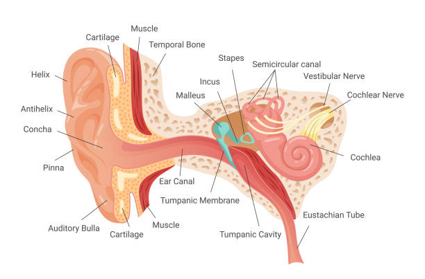
Ear and Their Functions :-
- The main of Ear is balancing of Human Body & other work is lesson / Collecting surrounding sound.
- The Pinna is responsible for collecting the sound in Human’s .
- Pinna’s muscle’s in Human ‘s is vestigial.
- Pinna is divided into two part’s
- Helix ( made up of elastic cartilage).
- Muscular Lobe.
Structure of Ear and Their functions :-
Human Ear are divided into Three part’s are following –
- Outer Ear ( Pinna , Ear Canal )
- Middle Ear ( Tympanium , Ossicle , Eustachian Tube )
- Inner Ear (Coachlea , 3 Semi-circular Canal’s)
- Stapes and Oval window are attached
Outer Ear and their functions :-

- Pinna :- It’s work is collecting sound waves and transfer to the Ear Canal. The degine of Pinna is in this to collect the maximum waves of their surrounding area.
- Ear Canal :-
- Hair :- The function of hair in Ear Canal is stop the foreign particle’s / dust particle’s that are present in Air.
- Wax (Cerumenous Glands) :- That is also work to trap the foreign particle’s / dust particle’s that are present in Air.
- The work of the Ear Canal is conduct the sound wave’s.
Middle Ear and their functions :-
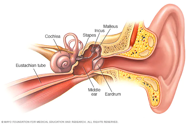
- Tympanum :- It is made up of “Connective Tissue”. It is vibrate’s when sound waves comes inside the Middle Ear.
- Eustachian Tube :- The Eustachian Tube connect Ear and Thrat and Pharynx. It is also maintain’s the air pressure balance occur’s tympanum.
- Ear Ossicle’s :- Smallest bones in body.
- (3 Bone’s in Each Ear)
- Ear Ossicles can done amplification of sound wave
- The 3 Bones of Ear Ossicle’s are following
- Malleus
- Incus
- Stapes
Malleus :- Malleus is the largest & First bone in these three bones. It look likes Hammer in shape.

It is formed by modification of Articulate Bone.
Incus :- Incus is the Middle bone in these three bones. It look likes Anvil in shape.

It is formed by modification of Quadrate Bone.
Stapes :- Stapes is the smallest & Last bone in these three bones. It look likes Stirrup in shape.

It is formed by modification of Hypo-Mandibular Bone.
Stapes is connected to cochlea through Oval(Ovalis) Window(Fenestra).
Window :-
They connect middle Ear ti Inner Ear. Their are two types are following :-
- Oval Window (Fenestra Ovalis) Ear and their functions
- Round Window (Fenestra Rotundus) Ear and their functions
1. Oval Window(Fenestra Ovalis) :-
The oval window, also known as the fenestra ovalis, is a connective tissue membrane located at the end of the middle ear and the beginning of the inner ear.

In the oval window the Scala vestibule (Perilymph fluid) are present
Helicotrema :- which connect the end part of , scala vestibuli and scalaa tympani.
“The Reissnar’s Membrane are present in the Oval Window at the lower side of the Oval Window“.
And Oval window open in Scala Vestibuli.
2. Round Window(Fenestra Rotundus) :-
The round window , is also known as fenestra rotundus, is also a connective tissue membrane located at the beginning of the inner ear and the end of the middle ear.
“The Basilar’s Membrane are present in the Round Window at the Upper side of the Round Window“.
The opening of Round Window in Scala Tympani. Ear and their functions
Note :- Ear and their functions Ear and their functions Ear and their functions
- On Basilar Membrane the Techorial membrane was present and the Sterocilia(When the Sterocilia cell’s move then impulse will be generated now this impulse will send to the Brain) was present on the Organ of Corti (it is a unit of Hearing).
- The Endolymph fluid was present between the Oval window and Round window.
Sterocilia → Movement → Impulse generated → Signal Send to Brain .
Inner Ear and their functions :-
In the inner ear, the sound waves are converted into electrical energy, which your hearing nerve delivers to your brain as sound, making it possible for you to hear. At the same time, your inner ear monitors your movements, alerting your brain to changes so your brain can let your body know what to do to stay balanced.
Coachlea :-
The cochlea is a fluid-filled, spiral-shaped cavity found in the inner ear that plays a vital role in the sense of hearing and participates in the process of auditory transduction. Sound waves are transduced into electrical impulses that the brain can interpret as individual sound frequencies. Ear and their functions
Mechansim of Hearing :-
- Amplified vibration enter’s into Scala vestibuli and they go from Scala vestibuli to Scala tympani because perilymph fluid also start vibrating but when perilymph fluid was vibrating then both Reissnar’s and Basilar membrane will also start vibrating.
- That vibrating conducted into scala media when vibrating enter’s into scala media then Endolymph fluid will also start vibrating.

- When Endolymph was start vibrating then the stereocilia was also start vibrating / moving. {Sterocilia is a cilia have a self movement generate. they can’t required any kind of external force for movement.}{Kinetocilia is a cilia they can’t required any kind of external force for movement. }
- Sterocilia can generate signal / impulse and that signal / impulse willl send to the brain by the help of auditory nerve (it is the 8th cranial nerve)(cranial nerve is sensory in nature.)
- Ear was located in temporal Bone.
Mechanism of Balancing :-
Responsible for balancing is Vestibular Apparatus.
Vestibular Apparatus
- 3 Semi – Circular Canal → Macula →Dynamic Equilibrium [Ampulla , Macula , Cristae Ampullaris.]
- Otoliths / Otaconia / Ear Duct → Saccule & Utricle → Static Equilibrium
 Structure of medula
- In Cupula the jelly like fluid are present an fluid can help in balancing (Dynamic Balancing.)
- Balancing jalantnus fluid was moving / vibrating then the stereocilia was also moving / vibrating and then generated signal and that signal was send to brain through neuron’s and the brain again send the signal to the muscle for movement.
- All semi-circular canal’s are perpendicular to each other.
Saccule And Utricle :-
- Stereocilia is responsible for static Equilibrium.
- When we are standing start then the fluid will be parallel when we start bending one side then the fluid come’s one side and the fluid apply the pressure on stereocilia and then the stereocilia generate impulse and that impulse send to the brin and the brain again send signal’s to the body part’s and muscle to maintain the body balance
- so the Saccule and utricle are responsible for maintain the body balance.