
Defintion of Heart :-
The hollow, muscular organ that pumps blood through the body of a vertebrate animal by contracting and relaxing. In humans and other mammals, it has four chambers, consisting of two atria and two ventricles.
External Structure of Heart :-

- The Atrium can obtain 1/3 part.
- The Ventricle can obtain 2/3 part .
- The Atrium and Ventricle are divided by Auriculo Ventricular Groove.
- The Upper side of the Heart (1/3 part) can be divided by Inter Auricular Sulcus / Septum.
- The Lower side (2/3 part) can be divided by Inter Ventricular Sulcus / Septum.

Protective Covering :-
It is Protected by Pericardium Layer.
- Peri mean’s outer side .
- Cardium mean’s heart .
- Portective sac of connective tissue.
- Filled with fluid.
- Pericardium layer made of connective tissue.
- Fluid Work :-
- Prevent from friction.
- Shock Absorbed.
- Prevent from Dehydration.
Internal Structure :-
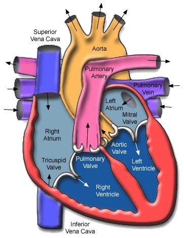
The De-oxygenated blood can be collected by superior Vena-cava into the Right Atrium and then blood can be transfer into Right Ventricle after full fill the Right Ventricular the blood can be transfer to the lung’s by the help of Pulmonary Artery and the blood can be oxygenated by the help of lung’s then the transfer blood again into the Left Atrium after full-fill the Left Atrium the blood goes to Left Ventricular chamber and after full fill Left Ventricular Blood can be transfer to the all body part’s by the help of Aorta.
Wall’s :-
Their are 3 layer’s of Heart
- Epi-cardium Layer
- Myocardium Layer
- Endo-cardium Layer

The wall’s are made up of
- Volentry Muscle’s
- Involentry Muscle’s
- Cardium Muscle’s
- The wall’s of Auricle’s is always thinner than the Ventricles
- Because Ventricles can pump the blood in the whole body part’s and the Auricle’s can collect the blood from all body.
- When we supply the blood to whole body then the pressure will be high then it is compulsory that the wall of Ventricles is thicker than the wall of Auricle’s to absorb the pressure.
- When we send blood to all body the we need more pressure do the wall’s of blood Ventricles thicker than the Auricle’s.
- The thickness of the wall always depend’s on the Myocardium layer of the Heart.
Fossa Ovalis :-
The Fossa Ovalis is a hole which is present after the birth.
Septum :-
The septum is the membrane which separate the right and left part of the heart . Separate De-oxygenated blood to Oxygenated blood.
Muscle’s of Heart :-
Ridge :-
Increase the surface area to hold blood in chamber’s.
- Small ridge in atria (Pectinate Muscle in Atria)
- Large ridge in Ventricle (Columnae Cornea/Trabeculae Cornea)
Chordea Tendineae :-
The chordae tendinae (CT) are strong, fibrous connections between the valve leaflets and the papillary muscles. These are attached to the leaflets on to the ventricular side and prevent the cusps from swinging back into the atrial cavity during systole.
Chordea made up off collagen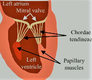
Papillary Muscles :-
There are 5 papillary muscles in the heart originating from the ventricular walls. These muscles attach to the tricuspid and mitral valve leaflets via the chordae tendineae and functionally prevent regurgitation of ventricular blood via tensile strength by preventing prolapse or inversion of the valves during systole.
Value’s :-
Values are muscular flapes . Flapes are prevent from flow of blood in last chamber of the heart.
- Tricuspid Valve. Located between the right atrium and the right ventricle.
- Pulmonary Valve(Pulmo-Semilunar Valve {P.S.L.V.}). Located between the right ventricle and the pulmonary artery.
- Mitral Valve / Bicuspid Valve. Located between the left atrium and the left ventricle.
- Aortic Valve (Aortic-Semilunar Valves{A.S.L.V.}).

Artery :- Artery they can take blood away from the Heart.
Vein / Venticle :- Vein / Ventrical they will collect the blood.
Structure of Heart :-

- The De-oxygenated Blood can be collected in Right Atrium after full fill the Right Atrium the blood can be transfer into Right Ventricle after full fill the Right Ventricle then the blood transfer into Lunge’s through the Pulmonary Artery
- The lunge’s can filter the blood by the help of Alveli
- In the Alveli the CO2 come out from Blood and O2 enter’s into the Blood this process is known as Gaseous Exchange
- After Gaseous Exchange the Blood come back again into the Heart in Left Atrium after the full fill the Left Atrium the Blood can transfer into Left ventricle
- After the full fill the Left Ventricle the blood can transfer into all body part’s through the Aorta.
Working Of Heart :-
- Our Heart is Myogenic {Myo – Muscle , Genic – Production}
- Our Cardiac System can generate our beat by own/itself. The impales is produced within the Heart.
- Our heart is Auto – Rythmicity.
- In Human Cardiac System the SAN(Sino-Auricular Node) and the AVN(Atrio-Ventricular Node)
- The SAN can take signal through Nerves from Brain and then the signal can transfer from SAN to A.V. Nodes and after that the electrical signal can transfer from A.V. Node to A.V. Bundles.
- From A.V. Bundles the electric impales can transfer into Left and Right Branch of A.V. Bundles.
- From Left to Right Branch of A.V. Bundles the electric impales can transfer into Purkinje Fibre’s.
S.A. Nodes :-
- S.A.N. is a pacemaker
- S.A.N can produce impales
- Both Left and Right Artium can contract together and both Left and Right Ventricle are also contract together.
- S.A. Node is Located in Right wall of Right Atrium.
A.V. Nodes :-
- A.V. Node is a Pacesetter.
- A.V. Node is Located in Left wall of Right Atrium.
- A.V. Nodes can re-amplify the impales and then the impales can transfer into A.V. Bundles.
- The A.V. Bundles are divided into two branches.
- Purkinje Fibre {One can make network of fibre}
- The action potential from the S.A. Node reach the Ventricles in the following path.
- Node ⇒ Atrial Muscles ⇒ A.V. Node ⇒ A.V. Bundle’s ⇒ Purkinji Fibre’s ⇒ Muscle’s of Ventricles (Wall of Ventricles)

Cardiac Cycle :-
Set of event’s which follow impales production by S.A. Node .
- Relax :- Diastole.
- Contraction :- Systole
- S.A. Node :(0.1 Second)- A.V. Node
- Joint Diastole :-(0.4 Second)


- Blood always flow from +ve pressure to -ve pressure {+ve means high pressure & -ve pressure }
- In joint diastole all the chamber are relaxed so inside the Heart -ve pressure will be present and blood collected / stored in the Heart
- T.V. & B.V. open & blood felling into auricles ⇒ Ventricle • 70% Ventricles are filled(Slow) at this time
Atrial Systole(0.1 second) :-
- After 0.4 second joint diastole the S.A. Node can generate impulse then the Auricles are contract and the blood can flow force fully into the Ventricles and the remaining (30%) part of the Ventricles are also filled with Blood.
- This can happened only in 0.1 second
- During this +ve pressure was present in auricle and -ve pressure present in Ventricles.
- This can be done Rapidly.
- When the Auricle are filling with Blood then that time the Ventricles will be relaxed.
Ventriclar Systole :-
- In the Ventricles +ve pressure will be present.
- I.V. and B.V. close.
- After filled with blood the blood can apply pressure P.S.L.U.(Semi-Lunar valves) and A.S.L.V. and the blood go outside through the A.S.L.V. to the body.
Blood Goes Out
- Aort. → (oxygenated) → Body
- Pulmonary Artery →(de-oxygenated) → Lunges
- When the blood go outside Ventricles then the Ventricles then the Ventricles are fully empty & then the T.V. & B.V. can suddenly open and due to this all blood of Auricles Chamber’s are collect that blood can full down into the ventricles.
Cardiac Output :-
- The volume of blood pumped out by each Ventricle , for each heartbeat , is known as the Stroke Volume.
- The Volume of blood pumped out by the heart from each Ventricle per minute is termed Cardiac output.
- Per beat = 70 ml
- Cardiac Output = S.V. × H.R.
- Cardiac Output = 70 × 72
- = 5040 ml/minute
- Athletes have the ability to increase the stroke volume and cardiac Output.
- The Cardiac Output generally increase in the active state and Decrease in resting condition.
Heart Beat :-
- Number of times it beat’s/mint
- Normal values = 70 – 80 times/mint
- Tachycardia = Heart Rate increase
- Bradycardia = Heart Rate decrease
Factors that Effect the Heart Beat :-
- Physical Activity
- Physical Condition & Health Condition.
- State of Mind
- Diet
- B.M.R.(Based Metaboli Rate)
Regulation of Heart Beat :-
- Nervous :- Medula Oblongata (Control)→ Cardiac centre (Send Message)→ S.A. Node
- Endocrine :-
- Acetylcholine → P.A.N.S( Parasentatic Nervous System) → decrease (Heart Rate)
- Thyroxin (Increase Heart Rate)
- Adrenalin
- Nor-Adrenalin
-
-
- Nor-Adrenalin and Adrenalin both are S.A.N.S. they Increase the Heart Rate
- P.A.N.S.(Parasentatic Nervous System) = They work in normal condition → Decrease Heart Reat
- S.A.N.S.(Sentatic Autonomic Nervous System) = They work in emergency condition.
-
Blood Pressure :-
Pressure exereted by blood on walls of blood vessels.
- B.P.(Blood Pressure) = S.P.(Systolic Pressure)/D.P.(Diastolic pressure)
-
- Normal Systolic Pressure = 120 mm/Hg
- Normal Diastolic pressure = 80 mm/Hg
- Normal B.P.(Blood Pressure) = 120/80 mm/Hg
- Hyper Tension (High B.P.) = 140/90 mm/Hg
- Hypo Tension (Low B.P.) = 100/60 mm/Hg
- Pulse Pressure = S.P. – D.P. ⇒ 120 – 80 =40mm/Hg
- Blood Pressure measured by Sphygmomanameter (SPO2)
- Heart Rate measured by Stethoscope
-
Blood Vessels :-
Artery :-
Which carry blood away from Heart and toward’s the organs.
- Tunica Externa made up of white fibrous connective tissue
- Tunica Media :- Smooth muscles

Vein :–
Which carry blood toward’s the Heart and collect from the organ’s & tissue’s.
- Tunica interna :- Squamous Epithelium

Capillary :-
- Tunica Interna & Tunica Media both are absent
- Tunica interna is present & it is also known as (Endothelium)

Lympth :-
- Blood – R.B.C.(Red Blood Cell’s) – Platelets
- Plasma + W.B.C.(White Blood Cell’s)
- Plasma + Lymphocyte + Monocytes
- Lympth is colourless.
- Proteins is also low.
- W.B.C. = 500 /nm3
- No Respiration Pigment
- Important Role in defence against disease.
- If lymph flow around tissue that , it is also known as Interstial Fluid / Tissue fluid.
- Acts as a middle man.
- Help in fat absorption.
Lymphatic Circulation :-
- Lymph Capillary are belind/close at one side.

Diapedsis :-
Squeezing / coming out of W.B.C. + Plasma.
- Heart → Aorta → Artery → Blood Capillary → Lymph vessels (Values more than veins) → Lymphatic Ducts (2 Lymphatic ducts are present at nack ression) → Sub Clavian veins (Right & Left) → Venacava → Heart.