
EAR :-
The engineering marvel because its sensory receptor can transduce sound vibrations with amplitude as small as diameter of atom of gold into electrical signal thousand times faster than photoreceptor can respond to light. It also contains receptor for equilibrium. It can also done Balance mechanisms of human body.
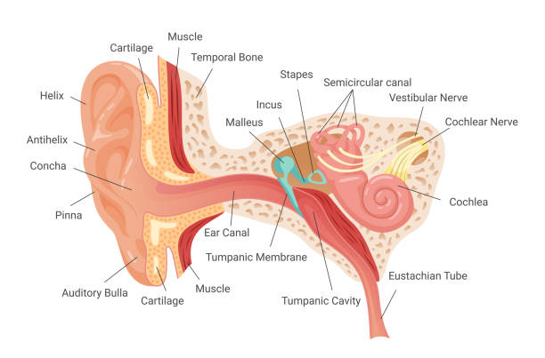
The Ear is divided into 3 main region :-

- External ear :- It collects sound waves and channel them inward.
- Middle ear :- It conveys sound waves to oval window.
- Internal ear :- It houses the receptor for equilibrium and hearing.
The external and outer ear consist of :–
a. Auricle
b. Auditory canal
c. Ear drum
Auricle (pinna) :-
Is a flap of elastic cartilage shaped like flared end of trumpet & covered by the skin
The rim of auricle is k/a helix and inferior portion is lobule
The lobule is highly blood supplied part of the ear
The auditory canal is & curved tube about 2.5 cm long
It is located in temporal bone & leads to ear drum
The tympanic membrane or ear drum is a thin semitransparent partition between the auditory canal and middle ear
The tympanic membrane is covered by epidermis and lined by simple cuboidal epithelium
Between the epithelial layer is connective tissue composed of collagen fibers and fibroblast
Near the exterior opening of auditory canal contains a few hair and specialized sweat glands known as cerumionous gland
Ceruminous gland secrets cerumen or wax
The combination of cerumen and hairs prevents dust and foreign object from entering the ear
Cerumen also prevent the damage to delicate skin of external ear canal by water and insects
Middle ear :-
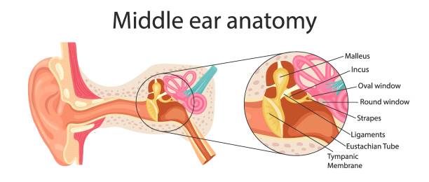
It is the smallest filled cavity in the petrous portion of temporal bone
It is lined by epithelium and separated by tympanic membrane from outer ear
It forms the internal ear by a thin bony partition that contains 2 membrane covered opening oval window & round window
3 smallest bone (Auditory ossicles) attached to it by ligaments which are connected by synovial joint
Auditory ossicles are :-
Malleus :-
It is a hammer shaped bone that attach to the internal surface of tympanic membrane (handle part). The head of malleus is articulate with the body of incus .
Inus :-
It is the anvil shaped bone and its head articulate with the head of stapes.
Stapes :-
It is stirrup shaped bone the footplate or base of stapes fits into oval window
Below the oval window there is another opening is round window which is enclosed by tympanic membrane .
2 skeletal muscles attach to the auditory ossicles :-
a) Tensor tympani :-
Supplied by mandibular branch of Trigeminal nerve (V)
It limits the movement and increase the tension on ear drum.
b) Stapedius :-
Supplied by the facial nerve (VII). It is smallest skeletal
Muscle. It protects the oval window and decrease sensitivity of hearing.
- Anatomy: The stapedius muscle is a tiny, cylindrical muscle located within the middle ear. It originates from the pyramidal eminence, a bony prominence on the posterior wall of the middle ear cavity, and inserts onto the neck of the stapes bone, one of the ossicles (small bones) of the middle ear.
- Innervation: The stapedius muscle is innervated by the facial nerve (cranial nerve VII). The motor fibers that control the stapedius muscle originate from the facial nerve nucleus in the brainstem and travel through the facial nerve before reaching the muscle.
- Function:
- Dampening Sound: The primary function of the stapedius muscle is to dampen excessive movement of the stapes bone in response to loud sounds. When activated, the stapedius muscle contracts and pulls the stapes bone away from the oval window, which connects the middle ear to the inner ear. This action reduces the transmission of sound vibrations to the inner ear, protecting it from damage caused by loud noises.
- Tuning: The stapedius muscle also plays a role in the process of sound localization and frequency tuning by adjusting the mechanical properties of the middle ear system.
- Reflex: The contraction of the stapedius muscle in response to loud sounds is known as the acoustic reflex or stapedial reflex. This reflex is a protective mechanism that helps prevent damage to the delicate structures of the inner ear from exposure to excessively loud noises.
- Clinical Significance:
- Hyperacusis: Dysfunction of the stapedius muscle can contribute to conditions like hyperacusis, where individuals experience increased sensitivity to normal environmental sounds.
- Acoustic Reflex Testing: The function of the stapedius muscle can be assessed through acoustic reflex testing, which measures the reflexive contraction of the muscle in response to loud sounds. This test is often used in audiological evaluations to assess hearing function and diagnose certain auditory disorders.
The anterior wall of middle ear contains an opening that leads to eustechian tube (pharyngotympanic)
It is consist of bot bone and elastic cartilage and connects the middle ear with nasopharynx
It is closed at the medial end (pharynx)
During swallowing and yawning its open and allow to air to enter or leave to middle ear until the pressure in the middle ear equal to atmospheric pressure
Inner ear :-
It is also known as labyrinth because of its complicated series of canal.it consist of 2 main division
a) Outer bony labyrinth
b) Inner membranous labyrinth
The bony labyrinth is series of cavities in the petrous portion of temporal bone. It is divided into 3 areas –
a) Semicircular canal :-
b) Vestibule:-
c) Cochlea :-
Semicircular anal and vestibule both the contains receptor of equilibrium and cochlea contains the receptor of hearing
The bony labyrinth is lined with periosteum and contains perilymph and chemically similar to CSF
Membranous labyrinth is a series of epithelial sac and tube inside the bony labyrinth. The epithelial membrane contains endlolymph
The level of K+ Ions is high for an ECF and play the important role to generate the electrical signal
a) Semicircular canal :-
superiorly and posteriorly from the vestibule there are 2 semicircular canal each of which approx. right angles to the outer 2
Based on their position they named as anterior, posterior and lateral semicircular canal. The anterior and posterior canals are vertical and lateral one is horizontal
At the one end of each canal is swallowen enlargement known as ampulla
The portion of membranous labyrinth lie inside the bony semicircular canal are known as semicircular ducts. These ducts connects with utricle and saccule
The vestibular branch of VIII CN consist of ampullary , utricular and saccular nerves and responsible for equilibrium
b) Vestibule :-
Is the oval central portion of the bony labyrinth. The membranous labyrinth in the vestibule consist of 2 sacs called utricle and saccule which are connected by a small duct.
c) Cochlea :-
is a snail shell bony spiral canal which is anterior to vestibule
It makes almost 3 turns around a central body core known as modiolus
Cochlea divided into 3 channels :–
a) Cochlear duct (scala media)
b) Scala vestibuli
c) Scala tympani
Cochlear duct is a continuation of membranous labyrinth into cochlea and filled with endolymph
The channel above the cochlear duct is scala vestibuli which ends at the oval window
The channel below is scala tymapni which ends at the round window
Both scala vestibuli and tympani are the part of bony labyrinth and Contains perilymph and both are completely separated to each other by scala media except helicoterma
Vestibular membranes separates the cochlear duct from scala tymapni
On the basilar membrane organ of Corti (Spiral Organ) Rests
Two groups of hair cells are presents-
a) Inner hair cells : arranged in single row
b) Outer hair cells : arranged in 3 layers
At the tip of hair cells 40-80 long hair like micro villi arranged known as stereo cilia
At the base end inner & outer hair cells synapse with VIII CN
A flexible gelatinous membrane cover the hair cells of spiral organ known as tectorial membrane. The body of hair cells rest on basilar membrane
PHYSIOLOGY OF HEARING :-
1. The auricles collects and directs the sound wave in auditory canal
2. Sound wave strike on tympanic membrane and the high & low pressure of air cause tympanic membrane to vibrate
3. The central area of ear drum connects to malleus which also start to vibrate. The vibrations is transmitted from the malleus to incus to stapes4. As stapes moves back & forth it pushes the membrane of oval window in & out
5. The oval window vibrates 20 times more vigorously than ear drum
6. The movement of oval window sets up fluid pressure waves in perilymph of cochlea
7. Oval window pushes on perilymph to scala vestibuli
8. Pressure waves are transmitted from scala vestibuli to scala tymapni to round window
9. Pressure wave push the vestibular membrane back & froth & creates the pressure waves in the endolymph inside the cochlear duct
10. The pressure wave in the endolymph causes the basilar membrane to vibrate
11. This moves the hair cells of spiral organ against tectorial membrane
12. This leads to bending of hair cells stercocilia which produce receptor potential that lead to generation of nerve impulse and leads to hearing
Pingback: Respiration (Part:-3) - online nursing point
Pingback: Eye and eye test (2024) - online nursing point
Pingback: Ear and their Functions (Part :- 2) - online nursing point